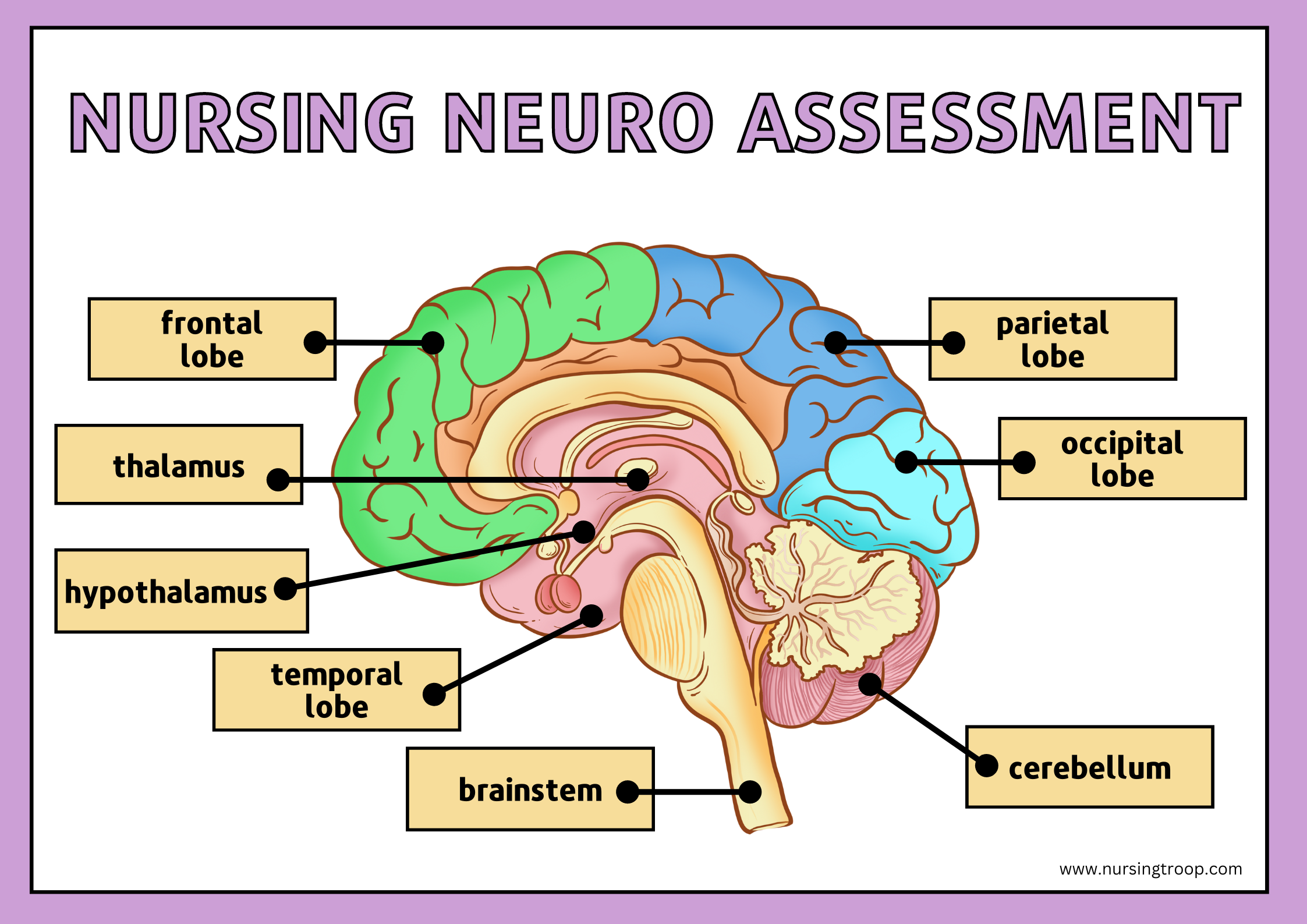
Doing a neuro assessment in nursing is critical to successful patient care and outcomes. Each situation requires different skills, techniques, and assessments depending on the severity of the illness or injury. In this blog post, we’ll cover all aspects of doing a proper nursing neuro assessment so you can be confident that you deliver the highest quality care possible for your patients. By walking through each step in detail, from pre-assessment preparation to documentation afterward, read on as we explore every aspect involved with successfully assessing neurological conditions.
Table of Contents
What is a Neurological Assessment?
A neurological exam is a physical examination of the nervous system. It looks for signs and symptoms indicating a disorder, injury, or disease of the brain, spinal cord, peripheral nerves, or muscles. Neurological exams evaluate mental and physical functioning in response to various stimuli, such as balance and coordination tests. They can also help diagnose diseases such as stroke, multiple sclerosis (MS), Alzheimer’s disease, cerebral palsy (CP), and other forms of dementia.
Who may need a Neurological Assessment?
A neurological exam may be needed for patients with a recent illness or injury, or those showing signs and symptoms of a nervous system disorder. Additionally, patients with a prior neurological condition or diagnosis should undergo routine evaluations to monitor their condition and track progress.
Why Neuro Checks Nursing Essential?
Neuro checks should always be in your arsenal of expertise as a nurse. The importance of timely and accurate neuro checks cannot be overstated, as they can mean the difference between life and death for your patient.
Regularly monitoring a patient’s neurological status is critical in assessing their level of consciousness, detecting any changes or abnormalities in their motor function, and identifying any potential problems in their brain function.
In short, mastering the art of neuro check can make you an invaluable member of your healthcare team and help ensure the best possible patient outcomes.
Who Conducts a Neurological Assessment?
Typically, a neurologist or other physician with specialized training in neurology conducts neurological exams. In some cases, specially trained nurses and physical therapists may also be qualified to conduct neurological assessments.
How Long does the Assessment Take?
The doctor will first talk to you and your child about her symptoms to get a good idea of which areas to concentrate on. After that part, the actual exam takes about 30 minutes.
What is Pre-Assessment Preparation?
Before beginning any assessment, preparing yourself by gathering pertinent information about the patient’s medical history is essential. This includes any current medications they are taking, prior illnesses/conditions they have had treated, a summary of any recent neurological assessments they may have had, and any relevant laboratory results.
Understanding the patient’s specific needs for the assessment is also essential. For example, if the patient has a known history of seizures or cerebral palsy, there might be tests that are more suitable for them than others. Understanding what type of testing will best serve their individual needs is essential.
Clinical Significance
A proper neurologic examination is essential to diagnose a disease as early as possible. With adequate practice, this examination can be conducted quickly and accurately. Abnormal results should be further investigated and referred to the appropriate specialists.
Seven categories of Neuro Assessment Nursing include:
- Mental status
- Evaluation of the cranial nerves:
- Motor system
- Depp Tendon Reflexes
- Sensory system
- Coordination
- Station & Gait
Mental Status Assessment
Testing cognition provides valuable information about thinking, memory, and emotional states. You may be asked to:
- Describe a recent event or activity
- Name common objects
- Remember and repeat a series of numbers
- Follow commands
Glasgow Coma Scale
The Glasgow Coma Scale (GCS) is a vital tool used in medical settings to assess a patient’s neurological function. This scale considers a patient’s motor response, verbal response, and eye-opening response.
The GCS is widely used in emergency rooms, as it is a quick and effective way to determine the severity of a patient’s brain injury. The scale ranges from 3 to 15, with 15 being the highest score indicating a patient’s normal neurological function.
- The eye-opening response is scored as spontaneous – 4
- opens to verbal command – 3
- opens to pain – 2, and no response – 1.
- The verbal response is scored as oriented – 5
- confused – 4
- inappropriate responses – 3
- incomprehensible sounds – 2, and no response – 1.
- The motor response is scored as obeys commands – 6
- spontaneous movement or localizes to painful stimuli – 5
- withdrawal from pain – 4
- abnormal flexion (decorticate) – 3
- the abnormal extension (decerebrate) – 2, and no response – 1.
The scores are added and classified as minor brain injury – 13 to 15 points, moderate brain injury– 9 to 12 points, and severe brain injury– 3 to 8 points.
The GCS assesses a patient’s initial neurological state and allows medical professionals to monitor the patient’s progress. It’s clear that the GCS plays a significant role in treating and caring for patients with brain injuries.
- Motor response – This assesses the patient’s muscle strength and movement. The examiner evaluates how well the individual can move different body parts, such as arms, legs, hands, feet, and head.
- Verbal response– This assesses the patient’s ability to communicate. The examiner typically asks questions that require a response to determine if the individual can understand and formulate verbal responses.
- Eye-opening response: This assesses the patient’s ability to open their eyes and respond to visual stimuli. The examiner will ask questions to gauge how alert the individual is, as well as if they can recognize people or objects in their environment.
Levels of Consciousness
Another way to assess your patient’s neurological status is to describe their level of consciousness, ranging from alert to comatose.
- Alert – This patient is awake and responsive to you and the environment.
- Example: Smith is sitting up in bed, texting with his granddaughter. When you ask him a question, he answers appropriately, and he follows all your commands.
- Confused – This patient is awake but is not entirely orientated to the environment.
- Example: Smith is sitting in bed but does not understand what is happening around him. When you ask him about her, he answers questions incorrectly and does not remember his granddaughter’s name.
- Somnolent – This patient is sleepy but can be aroused with some effort.
- Example: Smith is lying in bed, and he seems to be sound asleep. He responds slowly to your questions, but you can eventually get him to wake up.
- Lethargic – This patient is difficult to arouse and may drift back to sleep.
- Example: Smith lies in bed and does not respond to your questions or commands. You have to shout at him several times before he wakes up, but he quickly drifts back to sleep.
- Obtunded – This patient requires repeated stimulation to remain awake but is still somewhat responsive.
- Example: Smith is lying in bed and does not initially respond to your questions or commands. You have to shout at him several times before he wakes up, but then he remains awake for a few minutes before falling back asleep again.
- Stuporous – This patient requires intense stimulation but may remain aroused for short periods.
- Example: Smith is lying in bed and does not respond to your questions or commands, no matter how loud you shout at him. You need to engage in physical stimulation, such as shaking his body or slapping his face to get him to wake up, but then he quickly drifts back to sleep again.
- Comatose – This patient has no response to stimulation and is unarousable.
- Example: Smith is lying in bed and does not respond to your questions or commands no matter how loud you shout at him or how much physical stimulation you give him. He remains wholly unresponsive and unarousable.
It’s important to note that neurological assessment can provide valuable information about a patient’s condition and should be done as part of the overall medical evaluation. Attention to these details can help you identify issues requiring further investigation and diagnosis.
Evaluation of the cranial nerves:
Cranial nerves are pairs running directly from the brain to various body parts. It is essential to assess these nerves as they provide information about how well the patient’s brain is functioning. Examining these nerves includes testing functions like vision, hearing, smell, and taste. The twelve cranial nerves are:
- CN I (olfactory nerve) – Ask the patient to smell a familiar odor. In the hospital, some options you can usually get your hands on coffee and possibly food items with a distinctive smell, such as lemon and orange. Peppermint is another good choice if your hospital utilizes essential oils. If the patient can’t identify the odor or says, “I don’t smell anything,” this is a sign of neurological impairment.
- CN II (optic nerve) – Test the patient’s vision and visual fields by having them look at their finger or a penlight. Ask them to identify shapes, letters, numbers, and colors. Have them close one eye and assess each eye individually.
- CN III (oculomotor nerve) – Move the patient’s eyeballs up, down, left, and right to assess the coordination of movements between both eyes. Assess pupillary diameter in dim light with a flashlight or penlight while covering one eye at a time.
- CN IV (trochlear nerve) – Test the patient’s extraocular muscles by asking them to look up and side-to-side.
- CN V (trigeminal nerve) – Ask the patient to clench their teeth together and then check for symmetrical movement of both facial muscles. Test muscle strength by having them try to open their mouth against resistance.
- CN VII (facial nerve) – Ask the patient to raise their eyebrows, then smile, pucker their lips and blow out against your finger. Test facial sensation by running a cotton swab along the skin of the forehead and cheeks.
- CN VIII (acoustic nerve) – Test hearing by having the patient use earphones and repeat words or numbers at a reduced volume.
- CN IX (glossopharyngeal nerve) – Ask the patient to say their name and practice speaking in full sentences.
- CN X (vagus nerve) – Ask the patient to count backward from 10 or swallow a sip of water while you watch for normal tongue movement.
- CN XI (accessory nerve) – Test the patient’s shoulder shrug by having them lift their shoulders up and down.
- CN XII (hypoglossal nerve) – Ask the patient to stick out their tongue and move it side-to-side, up and down.
- a.Don’t forget visual fields by confrontation – vision is processed by 1/3 of the cerebral hemispheres.
- b. Check pupils and eye movements – don’t forget testing saccades as well as pursuits
- c. Facial strength is best tested by observing the patient for asymmetries during natural speech and for symmetry of eye blinks.
- d. Lower cranial nerves (IX-XII) only need to be tested if dysphagia and dysarthria are present.
Motor Function Assessment
The motor system is responsible for controlling the body’s voluntary movements. It is essential to assess this system as it can provide insight into neurological damage. Motor assessment includes testing strength, coordination, balance, speed of movement, and reflexes.
- To test strength: Ask the patient to perform specific tasks, such as gripping your hand or lifting their arms up against resistance.
- To test coordination: Have the patient walk heel-to-toe in a straight line or stand on one foot. To test balance: Ask the patient to stand with their feet together, and their eyes closed for 30 seconds or more without losing balance.
- To test the speed of movement: Have the patient quickly move from sitting to standing or rapidly tap different body areas with their fingers.
- To test reflexes: Tap different body areas with a hammer or reflex hammer and observe how the patient responds.
The motor system assessment is integral in determining the severity of a brain injury and ensuring appropriate treatment planning. The results of this exam will help medical professionals determine if physical therapy or other interventions are needed to improve functioning. Additionally, following up with regular assessments can help track progress over time.
Depp Tendon Reflexes
The reflexes are an essential part of the neurological assessment as they provide insight into the overall functioning of the nervous system. Reflex testing involves tapping or pressing on specific body areas while assessing the response from a particular muscle group. The results show how well signals are sent and received between different body parts and how quickly they respond in different situations.
Standard reflex tests include; pupil dilation, ankle jerk (also known as Achilles tendon), knee jerk, and finger/toe-curling. Other tests to assess reflexes may include the Babinski and the Hoffman signs. The results of these tests can provide important information about motor control and the functioning of peripheral nerves.
Reflex testing is essential to a neurological assessment as it can help reveal signs of nerve damage or dysfunction that may have gone unnoticed during other parts of the exam. Additionally, tracking changes in reflex responses over time can also help indicate if there has been any improvement or decline in neurological functioning since previous assessments.
Reflexes are described on a scale between zero to five, with normal reflexes as 2+.
The reflex scale is as follows:
- 0: No reflex in the muscle that’s being tested
- 1+: Diminished reflex
- 2+: Normal reflex
- 3+: Brisk reflex
- 4+: Clonus (repeated jerking of the muscle)
- 5+: Sustained clonus (prolonged jerking of the muscle)
Sensory System
The sensory system is responsible for processing environmental information through various sense organs. This information is then used to respond appropriately to external stimuli and guide behavior.
To assess the sensory system, a neurological exam includes testing of light touch, temperature sensation, vibration sense, pain perception, and proprioception (joint position). The examiner will use tools such as a cotton swab or tuning fork to check the response in different areas of the body.
- Focus sensory testing on the patient’s symptoms
- Sensory testing is purely subjective, so don’t over-interpret
- Check for sensory level on the back if a spinal cord lesion is suspected
- Touching nose with eyes closed – an excellent test of proprioception
- The Romberg test tests proprioception (peripheral nerves and dorsal columns), and is not a test of cerebellar function!
Coordination
Coordination is the ability to move muscles and body parts smoothly and precisely without overshooting or underperforming. Coordination testing generally includes tests of gross motor coordination (e.g., balance, agility), fine motor coordination (e.g., finger-thumb opposition), and sensory integration (e.g., writing with eyes closed).
During the neurological exam, coordination can be tested using tools such as Romberg’s test for static balance, tandem gait for dynamic balance, finger-to-nose test for fine motor coordination, and heel-to-toe test for trunk stability.
The results from these tests provide valuable information about the functioning of the central nervous system and can help determine if further evaluation is needed. Additionally, coordination tests can track changes in functioning over time and measure the effectiveness of interventions or treatments.
Station & Gait
The station and gait exam is integral to the neurological assessment as it provides insight into the patient’s ability to balance, walk, and move in general. This portion of the exam can help determine if there are any issues with coordination or strength in any muscles or joints.
Station: The station test involves assessing how well a person stands on their own two feet without support from anyone or anything else. The examiner will assess posture, balance reactions, and arm swings while walking.
Gait: During the gait exam, the examiner will observe how the patient walks across a room or hallway, noting any abnormalities such as limping or circumduction (shuffling). They may also ask them to turn, walk in the opposite direction, or walk around obstacles.
The results of these tests can provide valuable insight into the patient’s motor control and coordination. Abnormal results may indicate further evaluation is needed for the underlying cause of any issues detected. Additionally, tracking changes in gait over time can help measure progress with interventions or treatments.
Other Important Assessment
Autonomic Nervous System
The autonomic nervous system (ANS) regulates many vital functions, such as heart rate, breathing, sweating, digestion, and blood pressure. Examining the ANS is essential to assessing overall neurological functioning and can help indicate if any abnormalities may need further investigation.
Standard tests used to assess the ANS include; monitoring changes in heart rate while the patient holds their breath or stands up quickly, measuring skin temperature at different points on the body, assessing pupil dilation in a dark room, and checking for sweating when exposed to hot or cold stimuli.
Can you give a neurological exam to an infant?
Yes, newborns and infants have a unique series of reflexes that we can test, including:
- Blinking: your infant will close her eyes in response to bright lights
- Babinski reflex: as your infant’s foot is stroked, her toes will extend upward
- Crawling: if your infant is placed on her belly, she’ll make crawling motions
- Moro’s reflex: a quick change in your infant’s position will cause her to throw her arms outward, open her hands, and throw back her head
- Startle: a loud noise will cause your infant to extend and flex her arms while her hands remain closed in a fist
- Palmar and plantar grasp: her fingers or toes will curl around a finger placed in the area
Each one of these reflexes disappears at a certain age.
When do we get the results of the exam?
Right after the exam. Your child’s doctor will talk with you about the initial hypothesis, what the exam showed, and what your next steps should be. The exam may indicate that another test is needed, such as a blood test, an MRI, or a nerve conduction study. Your child’s doctor will be happy to answer any questions.
General Neurological Assessment
Assessing a patient’s overall neurological functioning does not usually necessitate the in-depth testing of each cranial nerve. Nonetheless, it is still essential to carry out critical evaluations that indicate whether the patient is experiencing any neurologic decline or improvement.
The critical components of a general neurological assessment are:
- Assess the level of consciousness (you’ll often use the GCS for this)
- Check for cranial nerve involvement
- Assess the motor and sensory responses in all four limbs
- Check pupillary light response
- Evaluate coordination with rapid movements such as finger-to-nose or heel-to-shin tests.
- Test reflexes, including those of the deep tendon
- Assess for Babinski sign (play an essential role in determining upper motor neuron function)
- Observe gait and balance when walking.
- Perform any other tests that are indicated if a specific lesion is suspected.
- Monitor vital signs, including temperature, pulse rate, blood pressure, and respiratory rate.
- Pay attention to skin color, tone, texture, and other physical characteristics.
Once a general neurological assessment is complete, your patient’s doctor will discuss the results with you and develop a plan of care based on the findings. This plan may include further tests or treatments to improve their condition. Additionally, if any abnormal results are found, it can alert doctors to potential problems that require further investigation. By performing regular neurological assessments on your patients, you can monitor their progress and ensure they get the best possible care.
National Institutes of Health Stroke Scale:
The National Institutes of Health Stroke Scale (NIHSS) is a tool used to assess the severity of a stroke at the time it occurs. It considers physical and neurological deficits, such as loss of movement in an arm or leg, altered level of consciousness, and language difficulties. The NIHSS consists of eleven items scored on a scale from 0-42. Higher scores indicate more severe strokes, and lower scores indicate less powerful strokes. The results from this test can help clinicians determine how best to treat patients and what interventions may be most beneficial for recovery.
Your neurological examination includes an assessment of your motor functions. The NIHSS comprises eleven components, each assessing certain abilities on a scale from 0 to 4. A score of 0 signals normal functioning in that particular ability, whereas higher scores point to some degree of impairment. Each item’s scores are added together to calculate a patient’s total NIHSS score; the maximum possible score is 42, and the lowest achievable result is 0.
| Score | Stroke severity |
| 0 | No stroke symptoms |
| 1–4 | Minor stroke |
| 5–15 | Moderate stroke |
| 16–20 | Moderate to severe stroke |
| 21–42 | Severe stroke |
Best Methods For Neurological Assessment Nursing
When assessing neurological function, paying attention to any changes in your patient’s condition over time is essential. Regular neurological assessments can provide insight into the effectiveness of treatments or help identify any potential issues that may need further investigation.
There are a few best practices neuro assessment nursing, you should keep in mind when performing a neurological assessment:
- Start with an overall physical examination and take note of any abnormalities
- Check for reflexes and muscle tone in all four limbs
- Perform cognitive evaluations such as language, memory, orientation, and executive functioning tests
- Observe gait and balance when walking
- Perform specific tests that are indicated if a particular lesion is suspected (e.g., pupillary light response, Babinski sign)
- Monitor vital signs such as temperature, pulse, blood pressure, and respiratory rate
- Pay attention to skin color, tone, texture, and other physical characteristics
- Record any changes over time and compare them with previous assessments for trends
- Use standardized scales such as the GCS or NIHSS to assess overall neurological function
- Educate your patient about their condition to ensure they understand their treatment plan.
By keeping these best practices in mind when assessing a patient’s neurological status, you can provide them with an accurate diagnosis and ensure they receive the best possible care.
Who is at risk for a neuro status change?
Patients at risk of a neurological change include those with pre-existing conditions such as:
- Stroke
- Traumatic brain injury
- Dementia
- Parkinson’s disease
- Alzheimer’s disease
- multiple sclerosis
- Patients who’ve had a carotid endarterectomy
- Patients with atrial fibrillation
- A patient with increased ICP
- Sodium imbalances, especially hyponatremia,
- A patient with an elevated BP
- Hypoglycemia Patient
- A patient with an infection
- Patients with pH imbalances
- other degenerative illnesses.
Patients with any of these conditions should be closely monitored for changes in their state that may indicate a decline in their neurological status.
Additionally, certain medications can cause confusion or other cognitive side effects, which could lead to a neurological change. It is essential to monitor patients taking these medications regularly to catch any potential changes early and minimize the impact on their health.
Finally, it is essential to remember that anyone can experience sudden changes in their neurologic state due to an acute illness or injury; this is why regular neurological assessments are vital to any patient’s care plan.
I hope this summary of a complex subject has provided clarity and boosted your confidence as you prepare to administer care at the patient’s bedside.. The key takeaways are:
- Obtain a baseline neurological assessment whenever possible.
- Ensure that assessments are conducted with the nurse from whom you have received the patient and with the nurse to whom you are transferring care.
- Don’t be afraid to seek a second opinion if necessary.
- If any abnormalities present themselves, review the chart for more information; sometimes, patients have pre-existing neurological issues not initially reported.
- Whenever changes occur, or concern arises, contact the medical doctor immediately.
Neuro Assessment Checklist With Example
A neurological examination is an important part of diagnosing and treating any condition involving the nervous system. It includes tests that measure muscle strength, reflexes, sensation, coordination, balance, and mental status. Here is a checklist to help guide you through a comprehensive neurological assessment:
- Mental Status: Ask patients questions about their name, date of birth, current time, place and chief complaint to assess orientation and recall.
- Cranial Nerves: Test the patient’s visual acuity, pupil reaction, extraocular movements, facial sensation and facial muscle strength.
- Motor Function: Assess muscle tone, power, and coordination throughout all four limbs.
- Reflexes: Test reflexes in the upper and lower extremities using a reflex hammer.
- Sensation: Evaluate the patient’s ability to feel light touch, pinprick, vibration and position sense throughout all four limbs.
- Gait: Observe the patient’s gait pattern while they are walking; assess for balance problems or any other abnormalities.
- Cerebellar function: Test the patient’s coordination of finger to nose and heel to the shin.
- Coordination: Ask the patient to perform tasks such as writing a sentence or drawing a clock face, which assesses their fine motor control.
- Vital Signs: Take crucial signs at intervals throughout the exam to monitor changes in blood pressure, heart rate, respiratory rate, and temperature.
By using this checklist to guide your neurological assessment, you can ensure that you provide the best possible care for your patient.
Example:
Mary Smith is a 64-year-old female presenting with complaints of confusion and disorientation. On physical examination, her vital signs were within normal limits, and her mental status was assessed as follows: she was alert but slightly confused regarding time and place; her memory was intact, but she had difficulty performing tasks such as writing a sentence or drawing a clock face.
Her cranial nerve exam revealed bilateral visual acuity of 20/20; pupils equal in size and reactive to light; extraocular movements were intact; the facial sensation was intact bilaterally; muscle strength was 5/5 throughout; reflexes were 2+ symmetric bilaterally. Her motor exam showed normal muscle tone and power in all 4 extremities with no coordination deficits noted. Her gait was normal, without any ataxia or balance problems.
In conclusion, Mary’s neurological examination revealed no significant abnormalities, and her condition likely stems from other causes unrelated to the nervous system. Further testing and evaluation should be pursued to determine a definitive diagnosis.
By conducting a thorough neurological assessment, you can gain important information regarding your patient’s condition that may help lead to an accurate diagnosis and appropriate treatment plan. It can be an invaluable tool for providing successful patient care when done correctly.
I hope this guide has helped help you better understand the neurological assessment process and how to conduct one confidently. Best of luck!
References
- https://my.clevelandclinic.org/health/diagnostics/22664-neurological-exam
- https://en.wikipedia.org/wiki/Neurological_examination
- https://www.ncbi.nlm.nih.gov/books/NBK557589/
- https://en.wikipedia.org/wiki/National_Institutes_of_Health_Stroke_Scale
Mrs. Marie Brown has been a registered nurse for over 25 years. She began her nursing career at a Level I Trauma Center in downtown Chicago, Illinois. There she worked in the Emergency Department and on the Surgical Intensive Care Unit. After several years, she moved to the Midwest and continued her nursing career in a critical care setting. For the last 10 years of her nursing career, Mrs. Brown worked as a flight nurse with an air ambulance service. During this time, she cared for patients throughout the United States.

